Clam Anatomy
Subject – Northern Quahog Mercinaria Mercinaria
Clams are bivalves meaning they have shells consisting of two halves, or valves.
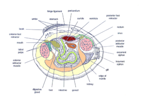
The valves are joined at the top, and the adductor muscles on each side hold the shell closed. If the adductor muscles are relaxed, the shell is pulled open by ligaments located on each side of the umbo.
The clam’s foot is used to dig down into the sand, and a pair of long siphons that extrude from the clam’s mantle out the side of the shell reach up to the water above (only the exit points for the siphons are shown).
Note: this image is colored to differentiate internal organs and are not the actual colors of the clam.
Clams are filter feeders. Water and food particles (and anything else that may be floating by) are drawn in through the incurrent siphon where tiny, hair-like cilia move the water to the gills. Food is caught in mucus produced by the gills. The gills also draw oxygen from the water flow.
From there, the cilia move the particles along food grooves toward the labial palps, where they are sorted. The labial palps secrete a mucus that entangles suspended food and nutrient particles within the water to produce a ball of food and mucus called a bolus. Afterwards, cilia on the palps direct the bolus into the mouth.
Other particles—such as silt or excess phytoplankton are dropped onto the surface of the mantle, where the clam eventually gets rid of them in mucous-coated balls.
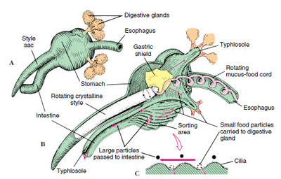
Food is processed by the digestive system which includes the stomach, intestines, rectum and anus.
The excurrent siphon carries away the water, disregarded (non-food) particles and waste.
Discarded waste material processed through the digestive system into the water column is called feces. Rejected material transported though the mantle is called pseudofeces.
Like all bivalves, scallops lack actual brains. Instead, their nervous system is controlled by three paired ganglia located at various points throughout their anatomy, the cerebral or cerebropleural ganglia, the pedal ganglia, and the visceral or parietovisceral ganglia.
 More about the shell:
The hard calm has its soft tissue surrounded by a shell consisting of two halves (valves).
When the clam is active and feeding, the shell continues to grow.
The shell consists of calcium carbonate and is held together by a though, pliable ligament at the top (dorsal) section of the animal called the hinge.
The hinge allows for the opening and closing of the shell. In close proximity to the hinge is the umbo commonly known as the “beak” The umbo is the oldest section of the shell with subsequent shell growth radiating out from it.
The shell is “spring loaded open” by the hinge. When alive, the two adductor muscles keep it tightly closed.
Nacreous Layer (nacre)
The innermost layer of the shell which touches the clam and connects it to the shell.
Also known as “Mother of Pearl”, this layer is smooth and made up of soothing material
Prismatic Layer
The middle layer of the shell which adds stability and strength to support the outer layer and the inner layer.
It neither touches the clam directly nor the outer elements of the water.
Periostracum Layer
The outermost layer of the shell and is extra hard as a protection against predators.
The main purpose of the outer layer is protection with a secondary purpose of supporting the clam inside.
It is what gives color to a mollusk’s shell.
Fun Fact: The word “periostracum” means “around the shell”
Inside the shell…
A clam has a mantle which surrounds its soft body.
A muscular foot which enables the clam to burrow itself in mud or sand.
The soft tissue above the foot is called the visceral mass and contains the clam’s body organs.
 The mantle
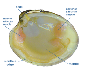
The mantle is a soft, retractable organ attached to both sections of the inside of a clam’s shell and is directly responsible for creating and growing the clam’s shell.
The mantle processes the calcium deposits that it has stored over weeks or months.
The clam opens its shell and extends its mantle to the edges of both sides.
The mantle releases the glue-like calcium compounds along the edges of the shell.
The functions of the mantle:
- Enclose and protect the internal organs.
- Removal of solids discarded by the labial palps
- A thickened rim of muscular tissue at the mantle edge deposit new shell material at the shell edge

